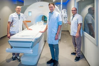
Sint-Trudo Hospital is one of the first hospitals in Flanders to introduce a photon-counting CT scanner in its radiology department. This new generation of CT technology reveals finer details within the body whilst using a lower radiation dose and less contrast fluid. Furthermore, it also provides spectral information.
Greater detail for an accurate assessment
A CT scanner uses X-rays to produce images of the inside of the body. Photon-counting CT records individual X-ray photons and their energy. This alternative method of measurement makes it possible to visualise small structures more sharply and to distinguish them more clearly from one another.

“The added value lies not only in sharper images, but above all in the information they provide us with,” explains Dr Kurt Buccauw, Head of the Radiology Department at Sint-Trudo Hospital. “With this technology, we can distinguish finer details and obtain spectral information. The sharper details help us to accurately assess abnormalities and support the treating doctor as effectively as possible. The spectral information, in turn, enables us to better define tissues and distinguish them from one another. We tailor each scan to the clinical need and the patient. In doing so, we aim for optimal image quality whilst minimising the radiation dose. The extent of this reduction in radiation dose depends on the type of scan, the patient and the selected settings. A lower dose does not, therefore, automatically apply to every scan.
Sharper images of various organs
For many imaging applications, photon-counting CT can provide sharper images, such as of the heart, lungs, ear, etc. One important application that deserves attention during ‘Heart Week’ is, for example, the examination of the coronary arteries – the blood vessels that supply the heart muscle with blood. Calcifications in these vessels can make it difficult to assess the severity of a narrowing. On a CT scan, these calcifications sometimes appear larger than they actually are. As a result, the narrowing may also appear more severe. Photon-counting CT can reduce this effect and thus helps to assess more accurately how much space remains in the blood vessel.
“For heart patients, what matters most is what we can do with that extra information,” says Dr Eva De Caluwé, a cardiologist at Sint-Trudo Hospital. “We want to know whether the coronary arteries are narrowed and how severe those narrowings are. The detailed images help us to assess this more accurately, even when there is significant calcification. They also result in fewer overestimates, which means that additional coronary imaging and the associated radiation exposure can be avoided. Our patients certainly benefit from this.

Expert care close to home
The fact that Sint-Trudo Hospital is one of the first hospitals in Flanders to invest in this new photon-counting CT is remarkable and demonstrates its full commitment to the further development of healthcare provision, and specifically to imaging within that field.
Less than two years ago, the hospital also commissioned one of the most powerful NMR machines, featuring a 3-Tesla magnet, which guarantees the highest-quality NMR images.
The DEXA scanner has been replaced, enabling radiologists to visualise total body composition with a minimal radiation dose, thanks to precise measurement of the ratio and distribution of bone mass, fat-free muscle mass and visceral fat throughout the body.
In addition, a new OPG unit has been given a central position in the department to enable dental and temporomandibular joint imaging to be carried out in accordance with the latest standards.
The SPECT-CT has also been replaced by a scanner that has been upgraded from 6 to 64 slices, drastically increasing image resolution. As a result, even subtle anatomical abnormalities and minor lesions can be detected much more quickly and accurately.